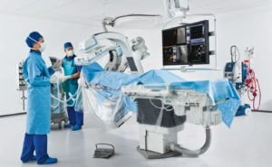
C-ARM Facility
The C-ARM facility is an essential part of modern medical diagnosis and treatment, providing real-time, high-quality imaging during various medical procedures. With its advanced imaging capabilities, C-ARM technology is widely used in orthopaedics, neurosurgery, trauma care, and other specialities. This article will delve into what C-ARM is, its applications, benefits, and how it plays a vital role in patient care. C-ARM Facility

What is C-ARM?
A C-ARM is a specialized medical imaging device that provides real-time, high-quality X-ray images during various medical procedures. Unlike traditional stationary X-ray machines, the C-ARM is portable and can be maneuvered around the patient during surgeries and diagnostic procedures. Its name comes from its C-shaped arm, which houses the X-ray source and detector, allowing for flexible movement and imaging from multiple angles.
In orthopaedic surgery, the C-ARM system allows for continuous imaging throughout the procedure, which is essential for ensuring precision during surgeries such as fracture repairs, joint replacements, and spinal surgeries. The ability to obtain immediate feedback during the operation aids in making real-time adjustments, ensuring that the procedure is performed with maximum accuracy.
Key Features of C-ARM Facility
Real-Time Imaging:
The primary advantage of the C-ARM is its ability to provide continuous, real-time X-ray imaging. This is particularly useful in surgeries where live imaging is essential for accurate placement of screws, plates, or other devices. The C-ARM allows surgeons to visualize the surgical site instantly, making it easier to adjust positioning and alignment during the procedure.Portability and Flexibility:
Unlike traditional X-ray machines that are fixed in place, the C-ARM is designed to be portable and flexible. This makes it ideal for use in the operating room, where it can be adjusted to various positions to get the best possible angle for imaging. Its compact design also allows it to fit in smaller spaces and be moved around the surgical table for better access to the patient.High-Resolution Imaging:
Modern C-ARM systems produce high-resolution, high-quality images that are essential for accurate diagnosis and treatment. The clarity of the images ensures that even the smallest fractures or misalignments are detected, allowing surgeons to take the necessary steps to correct any issues before completing the surgery.Minimal Radiation Exposure:
The C-ARM technology is designed to minimize the amount of radiation exposure to both the patient and medical personnel. With advanced filters, lower radiation doses, and digital imaging capabilities, C-ARM devices help to reduce the risks associated with traditional X-ray machines, ensuring a safer experience during procedures.Integration with Navigation Systems:
Many modern C-ARM systems can be integrated with surgical navigation systems, such as computer-assisted surgery (CAS). This combination provides even more precise control during procedures, enabling surgeons to plan the surgery in advance and monitor the exact placement of surgical instruments and implants in real-time.
Applications of C-ARM in Orthopaedic Surgery
Fracture Management:
C-ARM imaging is invaluable in the management of fractures, particularly complex fractures that require precise alignment during surgical fixation. Surgeons can use the C-ARM to guide the accurate placement of screws, plates, and rods, ensuring that fractures are properly set and stabilized.Joint Replacement Surgery:
In procedures like total hip replacement (THR) and total knee replacement (TKR), C-ARM systems provide crucial imaging that helps the surgeon properly align implants. Real-time feedback ensures that the prosthetics are positioned correctly, improving the longevity and function of the implants.Spinal Surgery:
For spinal surgeries, especially those involving complex procedures such as spinal fusion or the insertion of spinal screws, the precision offered by C-ARM imaging is crucial. Surgeons can ensure the correct placement of screws and other instruments by monitoring the surgery in real-time, reducing the risk of complications.Minimally Invasive Surgeries:
In minimally invasive procedures, such as arthroscopy and percutaneous screw insertion, the C-ARM is indispensable. The device provides continuous imaging guidance, allowing surgeons to work through smaller incisions while still obtaining precise information about the surgical site.Trauma Surgery:
Trauma cases often require quick decision-making and precise procedures. The use of C-ARM during trauma surgery allows orthopaedic surgeons to instantly assess the injury site and perform surgery with improved accuracy. This is especially useful in situations like fracture reduction and joint dislocations, where immediate visualization is key to achieving the best outcomes.
Applications of C-ARM Technology
C-ARM technology is used in a wide range of medical specialties, with its most common application in orthopaedic surgery, trauma care, and pain management. Some of the most common uses of C-ARM include:
1. Orthopaedic Surgeries
C-ARM is frequently used in orthopaedics to assist in joint surgeries, spinal procedures, and fracture management. For example, during joint replacement surgeries such as hip and knee replacements, C-ARM helps the surgeon position implants correctly and ensures the proper alignment of bones. It also helps in:
- Fixing fractures with pins, plates, or screws.
- Performing minimally invasive procedures, such as arthroscopy.
- Monitoring spinal alignment during spinal surgeries.
- Assisting in complex bone surgeries for better precision and less invasive approaches.
2. Trauma and Emergency Care
In trauma care, C-ARM provides real-time imaging for emergency procedures. It is used to evaluate fractures, dislocations, and injuries, especially when patients have multiple injuries that require immediate attention. C-ARM allows trauma specialists to perform the necessary interventions swiftly and accurately, improving the overall patient outcome.
3. Neurosurgery
In neurosurgery, C-ARM is used to guide the surgeon during delicate procedures, such as spinal surgery, brain tumour removal, or spinal cord injury repair. It provides real-time imaging to help doctors navigate around critical nerves and blood vessels, reducing the risk of complications.
4. Pain Management
C-ARM technology is also employed in the diagnosis and treatment of chronic pain. It is used to guide procedures such as:
- Epidural injections
- Nerve blocks
- Joint injections
By offering detailed imaging, C-ARM assists in delivering pain management therapies with high accuracy, increasing the effectiveness of these treatments and improving patient comfort.
5. Cardiovascular Procedures
In some cases, C-ARM is utilized in cardiovascular procedures, such as angiography or stent placements, to monitor blood flow and ensure correct placement of medical devices.
6. Gastrointestinal and Urological Procedures
C-ARM also plays a role in various diagnostic and therapeutic procedures within the gastrointestinal and urological fields, offering high-definition imaging for correct diagnosis and treatment.
Benefits of C-ARM Facility
The integration of C-ARM in clinical practice provides a number of advantages for both patients and healthcare providers:
1. Real-Time Imaging
One of the key benefits of C-ARM technology is its ability to provide real-time imaging during medical procedures. This allows surgeons to make immediate adjustments during the surgery based on live images, enhancing the accuracy of the intervention and minimizing risks.
2. Enhanced Precision
C-ARM allows for high-definition, high-quality images of the internal structures, which is especially critical during procedures that require extreme precision. The ability to view structures in real time helps surgeons maintain the correct position and alignment, reducing the chances of errors.
3. Minimally Invasive Procedures
The use of C-ARM in minimally invasive procedures, such as arthroscopy and endoscopy, has revolutionized the way certain surgeries are performed. C-ARM enables doctors to conduct surgeries through smaller incisions, resulting in less tissue damage, faster recovery times, and minimal scarring.
4. Mobile and Flexible
The C-ARM system is highly portable, making it ideal for use in operating rooms, emergency departments, and intensive care units (ICUs). It can be easily moved around and adjusted to accommodate different patient positions and procedural requirements.
5. Reduced Radiation Exposure
Modern C-ARM systems are designed to reduce the amount of radiation exposure to both the patient and healthcare providers. With the ability to adjust radiation doses and capture images at various angles, the C-ARM offers a safer option for patients undergoing imaging-guided procedures.
Conclusion
The C-ARM facility has become an indispensable tool in the medical field due to its ability to provide high-quality, real-time imaging for a wide range of medical procedures. From orthopaedics and neurosurgery to trauma care and pain management, C-ARM technology enhances the accuracy and precision of interventions, improving patient outcomes and reducing recovery times. Its mobility, flexibility, and the ability to reduce radiation exposure make it a valuable asset in modern medical practice, revolutionising the way surgeons and specialists approach complex surgeries.
